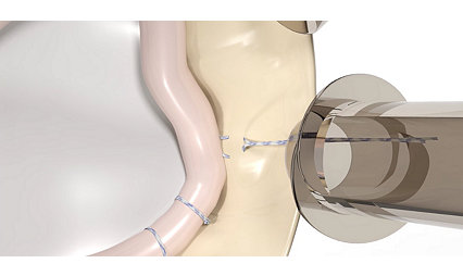
Iconix HA+
The future of all-suture.
Dual-modality HA+ coating
The Iconix HA+ coating was designed with HA and bioglass as these materials have been shown to accelerate bone healing at early implantation time.1,2,3
Hydroxyapatite + Bioglass
Hydroxyapatite is a stable compound shown to be osteoconductive and osteophyllic,4 with a close structural and chemical resemblance to bone material.5
Bioglass is a rapidly dissolving compound with two modes of bioactivity: bone bonding and osteostimulation.6,7,8
The Hydroxyapatite and bioglass have been shown in published studies to promote biological fixation between bone and a coated implant.1,9
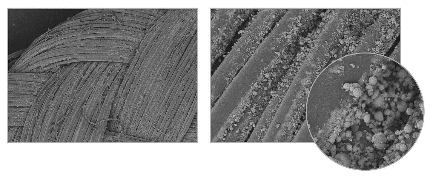
Osteostimulatory effect
Upon implantation, the ionic constituents of bioglass may be released into the surrounding environment and may react with bodily fluids to facilitate the deposition of a thin layer of physiologic calcium phosphate at its surface, thus attracting osteoblasts to the layer to create a matrix that promotes an osteostimulatory effect.6,7,8,10
In conjunction, the HA surface may act as a nucleating site for bone minerals,10,11 thus promoting the adhesion and proliferation of osteoblastic cells on the anchor surface.1,12
Creating the potential for accelerated bony integration
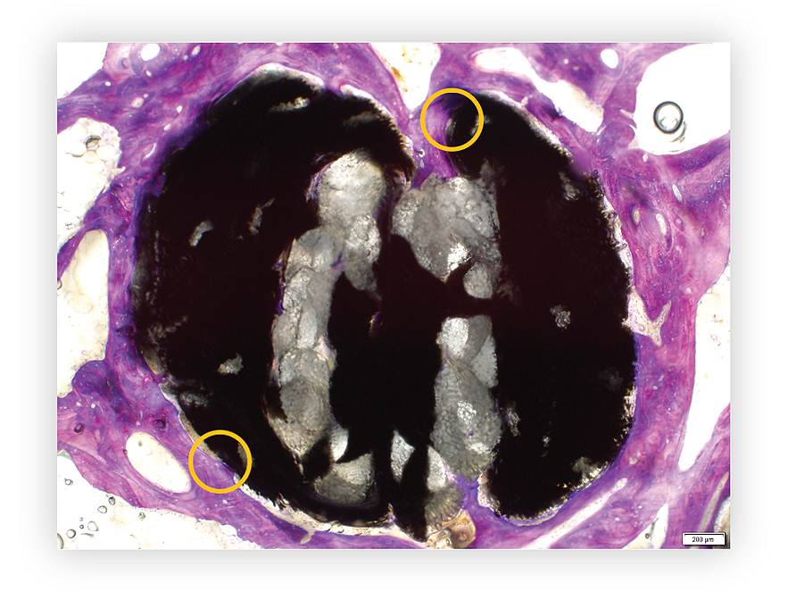
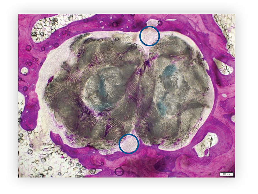
An ovine animal study, comparing Iconix HA+ to an uncoated all-suture implant, was designed to evaluate the implants with respect to bone ongrowth at 4, 8 and 12 weeks.In a blinded histomorphometry analysis at 8 weeks, the uncoated implant showed no integration (n=4), while Iconix HA+ showed bone integration in all samples (n=4).13
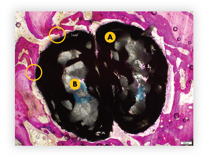
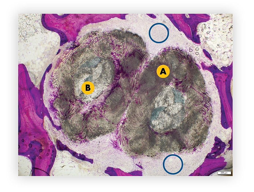
Comparison at 4 weeks
Histology images of an Iconix HA+ anchor (size 2.3mm) and a leading competitor all-suture anchor (single-loaded) at 4 weeks post-implantation in a large animal ovine study.13
A = Anchor
B = Suture
Gold circle = Bone formation
Blue circle = Voids between device and bone
Iconix HA+
Images show bone growing adjacent to the implant with some integration into the anchor.
Leading competitor all-suture anchor
Images show voids adjacent to bone.
Comparison at 12 weeks
Histology images of an Iconix HA+ anchor (size 2.3mm) and a leading competitor all-suture anchor (single-loaded) at 12 weeks post-implantation.13
Gold circle = Bone formation
Blue circle = Voids between device and bone
Iconix HA+
Leading competitor all-suture anchor
Backed by the Iconix family
The proven fixation, strength14 and versatility of the Iconix family now offered with HA+ coating.IntelliBraid technology
Targeted compression zones designed to create a bunching effect within the implant sheath for secure fixation with minimal bone removal
Self-centering technology
Iconix disposable drills have a unique self-centering technology to ensure accurate pilot hole placement*
Straight and curved guide options
The guide and obturator options allow for a variety of techniques.**
Indications
Indicated for surgical procedures in shoulder, hip, knee, foot and ankle, elbow and hand and wrist.**
* Applicable to PNs 3910500568 and 3910500571 ** See PUB-462 for full indications for use
References
1. Ielo I, Calabrese G, De Luca G, Conoci S. Recent Advances in Hydroxyapatite-Based Biocomposites for Bone Tissue Regeneration in Orthopedics. Int J Mol Sci. 2022 Aug 27;23(17):9721. doi: 10.3390/ijms23179721. PMID: 36077119; PMCID: PMC9456225.
2. Mohseni, E.; Zalnezhad, E.; Bushroa, A.R. Comparative investigation on the adhesion of hydroxyapatite coating on Ti–6Al–4Vimplant: A review paper. Int. J. Adhes. Adhes. 2014, 48, 238–257.
3. Arcos D, Vallet-Regí M. Substituted hydroxyapatite coatings of bone implants. J Mater Chem B. 2020 Mar 4;8(9):1781-1800. doi: 10.1039/c9tb02710f. PMID: 32065184; PMCID: PMC7116284.
4. Kurioka K, Umeda M, Teranobu O, Komori T. Effect of various properties of hydroxyapatite ceramics on osteoconduction and stability. Kobe J Med Sci. 1999 Aug;45(3-4):149-63. PMID: 10752309.Nanyang Technological University, Singapore.
5. J.L. Xu, K.A. Khor,5 - Plasma spraying for thermal barrier coatings: processes and applications,Editor(s): Huibin Xu, Hongbo Guo,In Woodhead Publishing Series in Metals and Surface Engineering,Thermal Barrier Coatings,Woodhead Publishing,2011,Pages 99-114.
6. J.R. Jones, D.S. Brauer, L.:G.D.C. HupaBioglass and Bioactive Glasses and Their Impact on HealthcareInt. J. Appl. Glass Sci., 7 (2016), pp. 423-434.
7. A. Hoppe, N. S. Gueldal, and A. R. Boccaccini, “A Review of the BiologicalResponse to Ionic Dissolution Products From Bioactive Glasses andGlass-Ceramics,” Biomaterials, 32 [11] 2757–2774 (2011).
8. Hench, L.L., Splinter, R.J., and Allen, W.C., Bonding Mechanisms at the Interface of Ceramic Prosthetic Materials. Journal of Biomedical Materials Research, 1971; 2(1): 117-141.
9. Chen Q, et al.Cellulose Nanocrystals--Bioactive Glass Hybrid Coating as Bone Substitutes by Electrophoretic Co-deposition: In Situ Control of Mineralization of Bioactive Glass and Enhancement of Osteoblastic Performance. ACS Appl Mater Interfaces. 2015 Nov 11;7(44):24715 25.
10. Amavedi S, Whittington AR, Goldstein AS. Calcium phosphate ceramics in bone tissue engineering: a review of properties and their influence on cell behavior. Acta Biomater. 2013;9:8037–45.
11. Bohner M, Lemaitre J. Can bioactivity be tested in vitro with SBF solution? Biomaterials. 2009;30:2175–9.
12. Mohseni, E.; Zalnezhad, E.; Bushroa, A.R. Comparative investigation on the adhesion of hydroxyapatite coating on Ti–6Al–4Vimplant: A review paper. Int. J. Adhes. Adhes. 2014, 48, 238–257.
13. Data on file at Riverpoint Medical - DNX0012 Animal Study - Iconix HA All- Suture Anchor.
14. DHFD15515
Related products and procedures
Shoulder Instability
Stryker’s 1.4mm shoulder instability anchor platform features the Iconix all-suture anchor and the NanoTack TT PEEK anchor, the smallest PEEK anchor on the market.
Learn moreIconix
Explore the comprehensive all-suture anchor platform, Iconix, with options for needle, self-punching, and tie-able anchor tape, designed to fit your orthopaedic sports medicine needs.
Learn more1000904581 rev A

