Airo TruCT in Proton Therapy
Interfraction imaging with Airo TruCT may enable treatment accuracy to support treatment fraction at the point-of-care. This allows for live updates to help improve the accuracy of the treatment as necessary. A change in patient position could mean that the target is not being fully treated or that healthy tissue structures are being unnecessarily subjected to proton radiation.
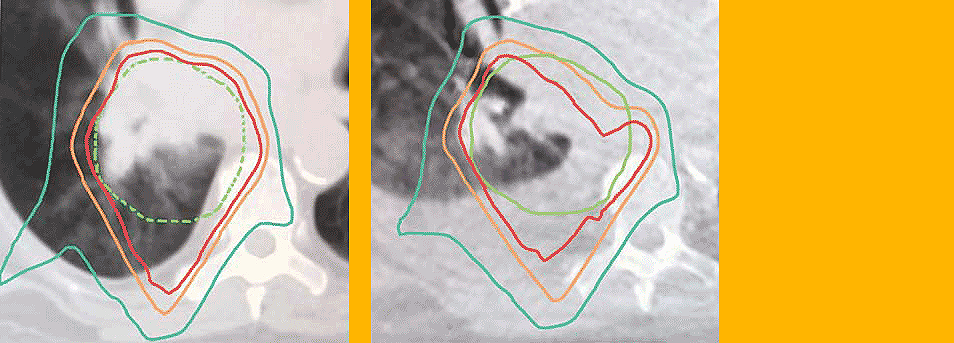
The presence of fluid and other soft tissue change can be readily visualized with helical CT imaging. Image sets with Hounsfield Units from Airo interfraction CT enable adaptive planning and treatment.

Left: Image of fluid accumulation in lungs (original)
Right: Image of fluid accumulation in lungs (3 weeks later)
Potential benefits in Proton Therapy
Portability
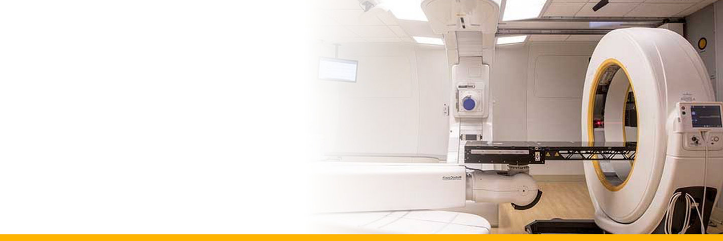
Airo’s self propelled transport mode and forward facing camera mean the system can easily be moved into and out of the Proton Therapy vault. It can also be moved between multiple vaults with little effort. Airo plugs into a standard wall power outlet.
Largest inner bore (107cm)
Airo's large inner bore enables the patient to be positioned and scanned all on the proton therapy treatment table. By minimizing patient movement during treatment, you are able to maintain higher positioning accuracy.
Reduced treatment time
Airo's ability to provide diagnostic quality imaging at point-of-care can help minimize the amount of time delay for patients requiring periodic interfraction CT imaging in Radiology.
TruCT-WB-1_26589

