Related categories
Software
CranialMap 3.0 Navigation Software is a simplified solution to incorporate navigation into neurosurgical procedures. From pre-op planning to intra-operative guidance and control, CranialMap 3.0 has been designed to meet the demands in today’s operating room and help deliver better patient outcomes.
Advanced imaging visualization enables you to compare unaffected and affected anatomy with automatic symmetrical visualization as a pre- and intraoperative guide.

Features and benefits
- Advanced auto segmentation capabilities for tumors, skin, brain, vasculature, ventricle and other volumes of interest
- Added features, including automatic image fusion between multiple CT, MR, CTA, MRA, fMRI and PET images with a single click
- Anatomical mirroring enables you to compare unaffected and affected anatomy with automatic symmetrical visualization as a pre and intraoperative guide
- CranialMask tracker offers a fully automated and simplified registration process and is the only tracker on the market that is completely sterile and designed for soft tissue placement
- 3D model enablement through custom and generic implant placement planning, modeling and verification
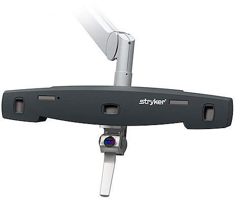
Stryker’s proprietary navigation camera
- Active optical technology
- Industry-leading accuracy for navigated procedures1
- Smart instruments with LED-based technology that enable wireless control of the software from the sterile field
CranialMask Tracker
The CranialMask Tracker offers a fully automated and simplified registration process.
- Only tracker on the market that is completely sterile and designed for soft tissue placement
- Small anatomical footprint and universal fit
- Enables pin-less workflow
- Full range head mobility
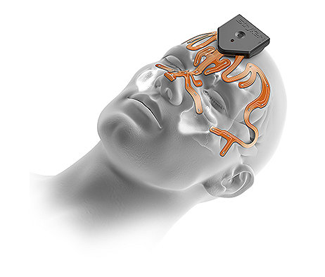
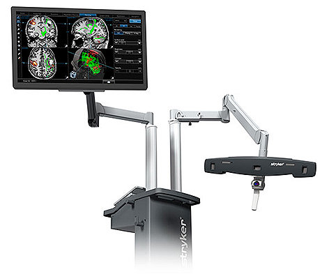
Flexibility to accommodate prone and lateral approaches
- Wide range of motion of camera arm to accommodate a variety of surgical procedures
- Built-in step-by-step workflow with mask registration and transfer to patient tracker
- Improved lateral visibility of mask for patient tracking
Advanced imaging visualization
- Import DTI/fMRI functional overlays and other advanced image sets
- Mirroring capabilities to overlay native anatomical images for reconstructive surgeries
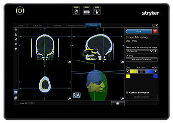
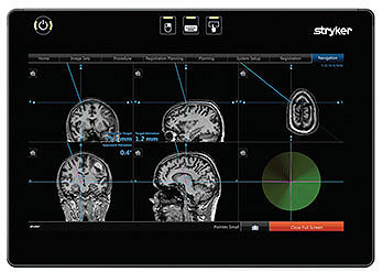
Customizable navigation views
- Easy-to-configure custom views based on surgeon’s preference
- Biopsy view with multiplane 3D and target views on one screen
Software that does more
- Image import via DICOM query/retrieve
- Microscope integration
- Frameless DBS capabilities with atlas options
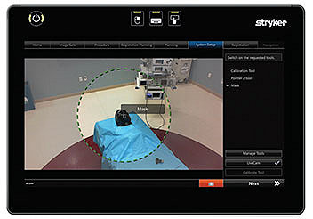
- Built-in LiveCam feature for simplified instrument setup
Smart instrumentation
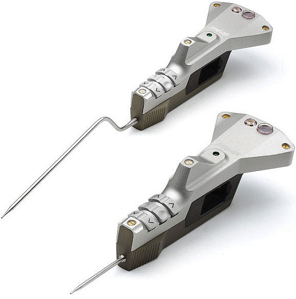
We’ve developed an array of LED-based instrumentation that integrates seamlessly with our CranialMap 3.0 Software. Wireless connections let you control the software remotely without compromising the sterile field. Convenient built-in LiveCam feature makes instrument setup easy.
- nGenius Universal Tracker
- Large pointer
- Small pointer
- Nasal pointer
- Shunt placement tool
- Left posterior-fossa pointer
- Right posterior-fossa pointer
- Vector calibration device
- Point calibration device
Additional accessories
- NavLock— 2-7 mm, 7-13 mm, 13-20 mm, 20-27 mm
- Universal joint screwdriver
- Tracker to Mayfield adapter
- Instrument battery
- Sterilization container
- Elfring R, de la Fuente M, Radermacher K. Assessment of optical localizer accuracy for computer-aided surgery systems; Comput Aided Surg. 2010;15(1-3):1-12.
Related categories
9100-004-797 Rev. None
